

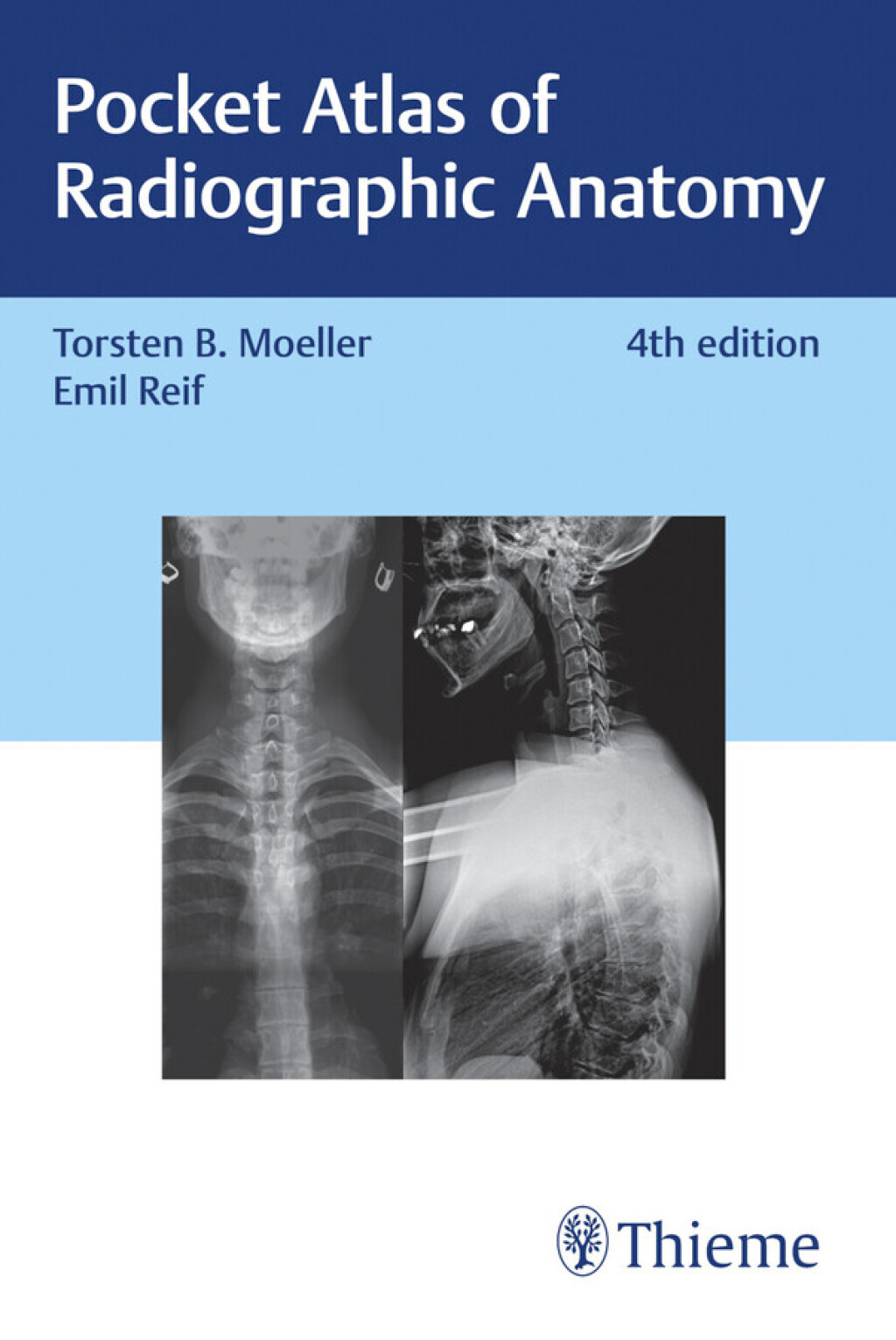

Pocket Atlas of Radiographic Anatomy 4th Edition – PDF/EPUB Version Downloadable
$49.99
Author(s): Torsten Bert Möller; Emil Reif
Publisher: Thieme Medical Publishers
ISBN: 9783132424418
Edition: 4th Edition
Radiographic anatomy at your fingertips
The Pocket Atlas of Radiographic Anatomy precisely describes the anatomical structures shown on conventional X-ray images and on the most important arthrographic and angiographic examinations.
The double-page spread format makes the book extremely user-friendly: The X-ray image is directly juxtaposed with a detailed drawing showing the clinically relevant anatomical structures. This enables simple, clear, and rapid identification and naming of the anatomical structures shown on the X-ray images.
Ideal for quick reference, the expanded 4th edition has been supplemented with new images, such as full-length views of the spine and leg, making it an indispensable resource for medical professionals and students alike.
Related products







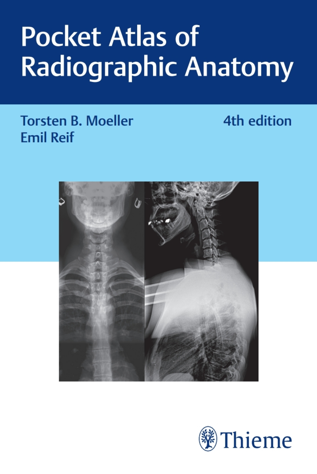

Pocket Atlas of Radiographic Anatomy 4th Edition – PDF/EPUB Version Downloadable
$49.99
Author(s): Torsten Bert Möller; Emil Reif
Publisher: Thieme Medical Publishers
ISBN: 9783132424418
Edition: 4th Edition
Radiographic anatomy at your fingertips
The Pocket Atlas of Radiographic Anatomy precisely describes the anatomical structures shown on conventional X-ray images and on the most important arthrographic and angiographic examinations.
The double-page spread format makes the book extremely user-friendly: The X-ray image is directly juxtaposed with a detailed drawing showing the clinically relevant anatomical structures. This enables simple, clear, and rapid identification and naming of the anatomical structures shown on the X-ray images.
Ideal for quick reference, the expanded 4th edition has been supplemented with new images, such as full-length views of the spine and leg, making it an indispensable resource for medical professionals and students alike.